You are here

leaf

Goldenrod leaf

I mentally restricted myself to blogging about tree leaves, but I've been watching for leaf miners since I started and haven't seen any until today and it was in a goldenrod leaf. So I'm writing about a goldenrod leaf (Fig. 1). The plant was near my bus stop and I only had a moment to grab the leaf before the bus came, so I didn't observe any other characteristics about the plant. I'm not sure it would have been possible to identify it more specifically until it flowers anyway. There are a lot of species of Solidago and only a few are really distinctive unless you know them pretty well.

Goldenrod Leaf MinerGoldenrod Leaf Miner

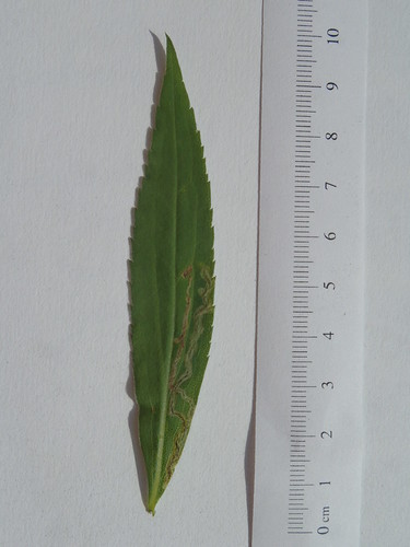
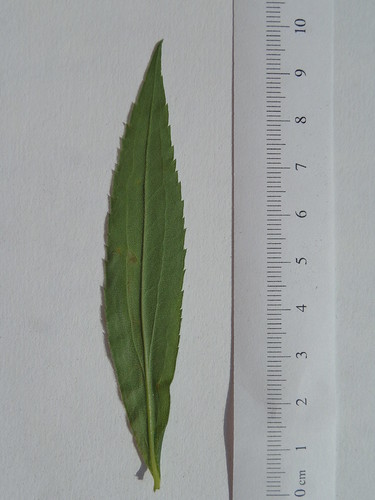
Figure 1. Goldenrod (Solidago sp.) leaf. Upper side is on left. Leaf miner damage visible.

Golderod leaves are lanceolate with a serrate margin. The leaf is light-green above and somewhat silvery underneath. The central vein protrudes under the leaf almost like a midrib.

The leaf miner track starts out around .2mm wide and leads toward the base of the leaf. When it reaches the intersection of a a smaller leaf vein with the middle vein, it turns abruptly, crosses the smaller vein, and heads toward the leaf margin. It runs along the leaf margin until it reaches the base of the leaf, where it reverses direction. The path returns along the leaf margin, but turns inward toward the middle and then meaders back and forth between the margin and the first major vein. There is a black trail inside much of the path, possibly frass or fecal material, The path leads to within a millimeter or two of the beginning and then stops.

Linden leaf

Near the Franklin Dining Commons Permaculture Garden are a line of European Linden trees (Tilia x europea). I noticed a branch where the leaves had been skeletonized by a small army of caterpillars (Fig 1.) The caterpillars were still active on one leaf and were lined up side-by-side eating along a single front on the underside of the leaf. I assume the underside because the cuticle is thinner there.

Tilia leafTilia leaf

Figure 1. Tilia x europea skeletonized by caterpillars. Upper side on left.

The Tilia leaf is heart-shaped with an asymmetrical base. The leaf margin is toothed, except at the base. The venation is palmate with 5 major veins that lead back to the leaf base.

Approximately three-quarters of the leaf area has been consumed by caterpillars leaving behind only the network of vascular tissue. In addition, I observed an aphid on the leaf which I have been unable to locate in the photographs. Perhaps it was on the underside of the stem while photographing was taking place.

Syringa reticulata leaf

A tree I love to hate is the Japanese Tree Lilac (Syringa reticulata). A leaf is illustrated in Figure 1. It is unobtrusive during most of the year, but in early June, it has large white flowers that smell like a scent they use for toilet paper.

Syringa reticulata leafSyringa reticulata leaf

Figure 1. Syringa reticulata leaf. Upper side is on left.

I started this post on Friday, but was too busy to complete it.

Prunus serrulata "Kwanzan" leaf

Near the "Fine Arts Center" bus stop where I get off near my office, there are two sakura trees that bloom later than the tree in my yard. I looked over the leaves and selected one (Fig. 1) to photograph and describe today.

Prunus serrulata "Kwanzan"Prunus serrulata "Kwanzan"

Figure 1. Prunus serrulata leaf. Upper side of leaf is on left.

I identified two differences between the Prunus serrulata "Kwanzan" leaf and the P. sargentii leaf I collected on July 5, 2017. The P. serrulata is flatter (less twisted) and the marginal teeth are all the same size and have a fine, drawn-out point with a black tip that the P. sargentii teeth lacked.

In other respects, the leaves are indistinguishable. The color, proportions, venation, and glands on the stem are roughly the same.

The surface of the leaf has some roughly linear marks. At first, I thought these might be leaf miners which look somewhat similar, but holding the leaf up to the light, I did not see any damage or change to the internal structures of the leaf. Other leaves had similar marks.

Prunus sargentii leaf

Today, I chose a leaf from a tree in my front yard. Figure 1 shows a leaf of Prunus sargentii, a Japanese Sakura tree. When this tree blooms (usually in April) the lush flowers last for only a few days, but are prized in Japanese culture as a symbol of the fleeting and transient nature of life.

Prunus sargentii leafPrunus sargentii leaf

Figure 1. Prunus sargentii leaf. Upper side of leaf is on left.

The leaves are ovate with a doubly-serrate margin and a left handed twist. The base is slightly asymmetrical. The venation is mostly alternate, but not entirely regular. Each vein curves at the end and connects with the next vein.

The stem is short and has a reddish pigment. There are two reddish glands on the stem the first about a millimeter behind the leaf and the second another millimeter behind the first.

There's no evidence of herbivory or damage to the leaf. I did observe some leaves that had parts missing, usually along the margins. Given the placement of the branches (above a driveway and a path used by people) its possible the leaves were damaged mechanically be people moving past the branches. I did observe a leaf hopper (Homoptera) on one leaf and some silk (spider? caterpillar?) on another.

Ulmus americana leaf

Since I started my leaf blog, I'm having a harder and harder time choosing a tree and leaf to look at—not because I'm running out of choices, but rather the opposite: there are so many tantalizing choices, its difficult to pick the next one. This time, I choose an American Elm (Ulmus americana) right behind the Morrill Science Center (Fig. 1).

Ulmus leafUlmus leaf

Figure 1. Ulmus americana leaf. Upper side is on left.

The Elm has ovate leaves with a double, or triple, serrate margin and asymmetrical base. The leaf has pinnate venation and there are approximately 13 side veins on each side of the leaf. About half of the side veins have branches near the margin of the leaf. The leaf has a very fine downy fuzz on both the upper and lower surfaces. The stem is quite short relative to the leaf.

Once again, I'm wondering a bit about the distance between the side veins and the sizes of the smallest areas of the leaf served by the venation system. The side veins seem close together, relative to other leaves I've looked at recently.

On the surface of the leaves are irregularly shaped galls with a pebbled surface. The galls have a yellowish color, although some have become black. On the underside of the leaf, there are gray fuzzy areas that correspond to the gall on the upper surface. There are a number of areas where the leaf surface is rough on the surface and fuzzy underneath, but where there is not an obvious gall: perhaps there are two phenomena.

Acer platanoides leaf

Today, I collected a leaf from a distinctive purple tree near the Franklin Dining Commons. The Norway maple (Acer platanoides) is a common ornamental tree that is often planted in cities because it is hardier and more tolerant of air pollution than native Sugar maples. It has two color varieties: the purple pigmented one and an unpigmented variety that's easy to confuse with Sugar maples. If you're trying to tell them apart, the bark is different on a Norway maple (a criss-cross diamond pattern) and Norway maples have a milky white sap.

Acer platanoides leafAcer platanoides leaf

Figure 1. Acer platanoides leaf. Upper side of leaf is on left.

The leaf is very dark green—almost black, in artificial light. The veins and stem are a reddish purple, especially on the underside of the leaf.

The leaf is palmately lobed with two small lobes near the base and 5 major lobes toward the tip. I had noticed yesterday that the stem of the Horse-chestnut seemed comparatively long, but this stem is much longer relative to the leaf. The stem is half-again longer than the leaf. I noticed, however, that there was a lot of variability in stem length.

The leaf has 11 holes where presumably insects have been consuming leaf material. Most holes run along a vein. One hole spans a vein in the leaf leaving just a narrow bridge across the hole. A number of holes have a kidney shaped pattern with smooth outward borders and a ragged inner border, perhaps related to the pattern of consuming the leaf.

On the underside of the leaf is a small, white cocoon. The area is covered over with a dense layer of white silk.The silk pulls the leaf together to make a small, bent section, where the pupa is concealed.

Aesculus leaf

I actually collected this leaf (Figure 1) yesterday while I was carrying the Coffee Tree leaf back. The Horse-chestnut one of my favorite trees in the spring when it flowers. People say the flowers look like candles and, on a dark, rainy day, the tree does seem to light up when the flowers are in bloom.

Aesculus leafAesculus leaf

Figure 1. Aesculus leaf. Upper side is on left.

The leaf is palmately compound with 7 leaflets. The leaves are ovate with irregularly toothed margins. The venation is not opposite, but nearly so. The stem seems extremely long, relative to the leaf.

The leaf has several patches of what appears to be a fungus. In each area is a brownish red region surrounded by a brownish region. The regions cross side veins, but rarely cross the mid-vein. There is some brownish material on the underside of the leaf where leaflets come together.

Gymnocladus leaf

The trees on the UMass Amherst campus are named the Frank A Waugh Arboretum and the link leads to an ArcGIS application that shows the location and identification of every tree (as of 2014). This is a great resource for being able to identify species, especially the ones that are not native to the region, like the Kentucky Coffee Tree.

Gymnocladus leafGymnocladus leaf

Figure 1. Gymnocladus leaf. Upper side of leaf is on left.

The main characteristic I use to recognize a coffee tree is that the leaflets are rotated away from the plane of the leaf itself. Most compound leaves appear oriented in a single plane.

This leaf is bi-pinnately compound. The first two branches are single leaflets, then two with ~8 leaflets, then branches with more (11 and 13). The branches are not evenly distributed: some are almost opposite, the last two are opposite. The leaflets, however are alternate.

The leaf appears almost pristine. There are several small areas of damage, the look to be mechanical damage of the leaf. There doesn't appear to be any evidence of phytophagy.

The stem has a waxy coating that wiped off when I carried the stem by the end.

Quercus Leaf

On Sunday, I collected an oak leaf. This is from a small oak tree in my yard, probably a red or black oak.

Quercus leafQuercus Leaf

Figure 1. Quercus leaf. Upper side of leaf is on left.

The leaves were rather diverse. Some were highly dissected, others broad and regular. Many of the leaves had big chunks eaten away. In some, the veins were left behind along with frass or detritus from whatever had consumed them. I remember one time at night, I saw small June beetles eating oak leaves, so I've always imagined that damage to oak leaves is caused by June beetles. It would be interesting to go back at night sometime and try to observe what causes the damage. I didn't see anything on the tree during the daytime (like a caterpillar) that looked like it might be causing the damage.

I migrated the Betula leaf blog entry to here from the class blog in Blackboard. I've posted this one here first to see whether it's easier to migrate one way or the other. Because Blackboard sucks.

Pages

Subscribe to RSS - leaf